Pathway and Gene Set Overdispersion Analysis
In this vignette, we show you how to use pagoda routines in the scde package to characterize aspects of transcriptional heterogeneity in populations of single cells.
The pagoda routines implemented in the scde resolves multiple, potentially overlapping aspects of transcriptional heterogeneity by identifying known pathways or novel gene sets that show significant excess of coordinated variability among the measured cells. Briefly, cell-specific error models derived from scde are used to estimate residual gene expression variance, and identify pathways and gene sets that exhibit statistically significant excess of coordinated variability (overdispersion). pagoda can be used to effectively recover known subpopulations and discover putative new subpopulations and their corresponding functional characteristics in single-cell samples. For more information, please refer to the original manuscript by Fan et al..
Preparing data
The analysis starts with a matrix of read counts. Here, we use the read count table and cell group annotations from Pollen et al. can be loaded using the data("pollen") call. Some additional filters are also applied.
data(pollen)
# remove poor cells and genes
cd <- clean.counts(pollen)
# check the final dimensions of the read count matrix
dim(cd)
## [1] 11310 64
Next, we'll translate group and sample source data from Pollen et al. into color codes. These will be used later to compare Pollen et al.'s derived annotation with subpopulations identified by pagoda:
x <- gsub("^Hi_(.*)_.*", "\\1", colnames(cd))
l2cols <- c("coral4", "olivedrab3", "skyblue2", "slateblue3")[as.integer(factor(x, levels = c("NPC", "GW16", "GW21", "GW21+3")))]
Fitting error models
Next, we'll construct error models for individual cells. Here, we use k-nearest neighbor model fitting procedure implemented by knn.error.models() method. This is a relatively noisy dataset (non-UMI), so we raise the min.count.threshold to 2 (minimum number of reads for the gene to be initially classified as a non-failed measurement), requiring at least 5 non-failed measurements per gene. We're providing a rough guess to the complexity of the population, by fitting the error models based on 1/4 of most similar cells (i.e. guessing there might be ~4 subpopulations).
Note this step takes a considerable amount of time unless multiple cores are used. We highly recommend use of multiple cores. You can check the number of available cores available using detectCores().
# EVALUATION NOT NEEDED
knn <- knn.error.models(cd, k = ncol(cd)/4, n.cores = 1, min.count.threshold = 2, min.nonfailed = 5, max.model.plots = 10)
For the purposes of this vignette, the model has been precomputed and can simply be loaded.
data(knn)The fitting process above wrote out cell.models.pdf file in the current directory showing model fits for the first 10 cells (see max.model.plots argument). The fitting process above wrote out cell.models.pdf file in the current directory showing model fits for the first 10 cells (see max.model.plots argument). Here's an example of such plot:

The two scatter plots on the left show observed (in a given cell) vs. expected (from k similar cells) expression magnitudes for each gene that is being used for model fitting. The second (from the left) scatter plot shows genes belonging to the drop-out component in red. The black dashed lines show 95% confidence band for the amplified genes (the grey dashed lines show confidence band for an alternative constant-theta model). The third plot shows drop-out probability as a function of magnitude, and the fourth plot shows negative binomial theta local regression fit as a function of magnitude (for the amplified component).
Normalizing variance
In order to accurately quantify excess variance or overdispersion, we must normalize out expected levels of technical and intrinsic biological noise. Briefly, variance of the NB/Poisson mixture processes derived from the error modeling step are modeled as a chi-squared distribution using adjusted degrees of freedom and observation weights based on the drop-out probability of a given gene. Here, we normalize variance, trimming 3 most extreme cells and limiting maximum adjusted variance to 5.
varinfo <- pagoda.varnorm(knn, counts = cd, trim = 3/ncol(cd), max.adj.var = 5, n.cores = 1, plot = TRUE)
The plot on the left shows coefficient of variance squared (on log10 scale) as a function of expression magnitude (log10 FPM). The red line shows local regression model for the genome-wide average dependency. The plot on the right shows adjusted variance (derived based on chi-squared probability of observed/genomewide expected ratio for each gene, with degrees of freedom adjusted for each gene). The adjusted variance of 1 means that a given gene exhibits as much variance as expected for a gene of such population average expression magnitude. Genes with high adjusted variance are overdispersed within the measured population and most likely show subpopulation-specific expression:
# list top overdispersed genes
sort(varinfo$arv, decreasing = TRUE)[1:10]
## DCX EGR1 FOS IGFBPL1 MALAT1 MEF2C STMN2 TOP2A
## 5.000000 5.000000 5.000000 5.000000 5.000000 5.000000 5.000000 5.000000
## BCL11A SOX4
## 4.755811 4.522795
Controlling for sequencing depth
Even with all the corrections, sequencing depth or gene coverage is typically still a major aspects of variability. In most studies, we would want to control for that as a technical artifact (exceptions are cell mixtures where subtypes significantly differ in the amount of total mRNA). Below we will control for the gene coverage (estimated as a number of genes with non-zero magnitude per cell) and normalize out that aspect of cell heterogeneity:
varinfo <- pagoda.subtract.aspect(varinfo, colSums(cd[, rownames(knn)]>0))Evaluate overdispersion of pre-defined gene sets
In order to detect significant aspects of heterogeneity across the population of single cells, 'pagoda' identifies pathways and gene sets that exhibit statistically significant excess of coordinated variability. Specifically, for each gene set, we tested whether the amount of variance explained by the first principal component significantly exceed the background expectation. We can test both pre-defined gene sets as well as 'de novo' gene sets whose expression profiles are well-correlated within the given dataset.
For pre-defined gene sets, we'll use GO annotations. For the purposes of this vignette, in order to make calculations faster, we will only consider the first 100 GO terms plus a few that we care about. Additional tutorials on how to create and use your own gene sets can be found in a separate tutorial.
library(org.Hs.eg.db)
# translate gene names to ids
ids <- unlist(lapply(mget(rownames(cd), org.Hs.egALIAS2EG, ifnotfound = NA), function(x) x[1]))
rids <- names(ids); names(rids) <- ids
# convert GO lists from ids to gene names
gos.interest <- unique(c(ls(org.Hs.egGO2ALLEGS)[1:100],"GO:0022008","GO:0048699", "GO:0000280", "GO:0007067"))
go.env <- lapply(mget(gos.interest, org.Hs.egGO2ALLEGS), function(x) as.character(na.omit(rids[x])))
go.env <- clean.gos(go.env) # remove GOs with too few or too many genes
go.env <- list2env(go.env) # convert to an environment
Now, we can calculate weighted first principal component magnitudes for each GO gene set in the provided environment.
pwpca <- pagoda.pathway.wPCA(varinfo, go.env, n.components = 1, n.cores = 1)We can now evaluate the statistical significance of the observed overdispersion for each GO gene set.
df <- pagoda.top.aspects(pwpca, return.table = TRUE, plot = TRUE, z.score = 1.96)
Each point on the plot shows the PC1 variance (lambda1) magnitude (normalized by set size) as a function of set size. The red lines show expected (solid) and 95% upper bound (dashed) magnitudes based on the Tracey-Widom model.
head(df)## name npc n score z adj.z sh.z adj.sh.z
## 57 GO:0048699 1 864 2.261079 27.019091 26.894993 NA NA
## 56 GO:0022008 1 911 2.233116 27.062973 26.913370 NA NA
## 30 GO:0000226 1 302 1.665067 11.226626 10.989344 NA NA
## 55 GO:0007067 1 330 1.626072 11.035135 10.814193 NA NA
## 37 GO:0000280 1 395 1.617566 11.650728 11.397103 NA NA
## 9 GO:0000070 1 96 1.512958 5.888795 5.555273 NA NA
- The z column gives the Z-score of pathway over-dispersion relative to the genome-wide model (Z-score of 1.96 corresponds to P-value of 5%, etc.).
- "z.adj" column shows the Z-score adjusted for multiple hypothesis (using Benjamini-Hochberg correction).
- "score" gives observed/expected variance ratio
- "sh.z" and "adj.sh.z" columns give the raw and adjusted Z-scores of "pathway cohesion", which compares the observed PC1 magnitude to the magnitudes obtained when the observations for each gene are randomized with respect to cells. When such Z-score is high (e.g. for GO:0008009) then multiple genes within the pathway contribute to the coordinated pattern.
Evaluate overdispersion of 'de novo' gene sets
We can also test 'de novo' gene sets whose expression profiles are well-correlated within the given dataset. The following procedure will determine 'de novo' gene clusters in the data, and build a background model for the expectation of the gene cluster weighted principal component magnitudes. Note the higher trim values for the clusters, as we want to avoid clusters that are formed by outlier cells.
clpca <- pagoda.gene.clusters(varinfo, trim = 7.1/ncol(varinfo$mat), n.clusters = 50, n.cores = 1, plot = TRUE)
The plot above shows background distribution of the first principal component (PC1) variance (lambda1) magnitude. The blue scatterplot on the left shows lambda1 magnitude vs. cluster size for clusters determined based on randomly-generated matrices of the same size. The black circles show top cluster in each simulation. The red lines show expected magnitude and 95% confidence interval based on Tracy-Widom distribution. The right plot shows extreme value distribution fit of residual cluster PC1 variance magnitude relative to the Gumbel (extreme value) distribution.
Now the set of top aspects can be recalculated taking these de novo gene clusters into account:
df <- pagoda.top.aspects(pwpca, clpca, return.table = TRUE, plot = TRUE, z.score = 1.96)
head(df)
## name npc n score z adj.z sh.z adj.sh.z
## 65 geneCluster.8 1 307 3.235994 12.803995 12.496650 NA NA
## 57 GO:0048699 1 864 2.261079 27.019091 26.894993 NA NA
## 56 GO:0022008 1 911 2.233116 27.062973 26.913370 NA NA
## 30 GO:0000226 1 302 1.665067 11.226626 10.989344 NA NA
## 72 geneCluster.15 1 287 1.642582 6.453696 5.947129 NA NA
## 55 GO:0007067 1 330 1.626072 11.035135 10.814193 NA NA

The gene clusters and their corresponding model expected value and 95% upper bound are shown in green.
Visualize significant aspects of heterogeneity
To view top heterogeneity aspects, we will first obtain information on all the significant aspects of transcriptional heterogeneity. We will also determine the overall cell clustering based on this full information:
# get full info on the top aspects
tam <- pagoda.top.aspects(pwpca, clpca, n.cells = NULL, z.score = qnorm(0.01/2, lower.tail = FALSE))
# determine overall cell clustering
hc <- pagoda.cluster.cells(tam, varinfo)
Next, we will reduce redundant aspects in two steps. First we will combine pathways that are driven by the same sets of genes:
tamr <- pagoda.reduce.loading.redundancy(tam, pwpca, clpca)In the second step we will combine aspects that show similar patterns (i.e. separate the same sets of cells). Here we will plot the cells using the overall cell clustering determined above:
tamr2 <- pagoda.reduce.redundancy(tamr, distance.threshold = 0.9, plot = TRUE, cell.clustering = hc, labRow = NA, labCol = NA, box = TRUE, margins = c(0.5, 0.5), trim = 0)
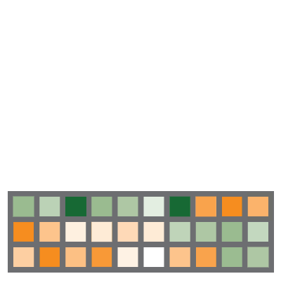
In the plot above, the columns are cells, rows are different significant aspects, clustered by their similarity pattern.The green-to-orange color scheme shows low-to-high weighted PCA scores (aspect patterns), where generally orange indicates higher expression. Blocks of color on the left margin show which aspects have been combined by the command above. Here the number of resulting aspects is relatively small. "top" argument (i.e. top = 10) can be used to limit further analysis to top N aspects.
We will view the top aspects, clustering them by pattern similarity (note, to view aspects in the order of increasing lambda1 magnitude, use row.clustering = NA).
col.cols <- rbind(groups = cutree(hc, 3))
pagoda.view.aspects(tamr2, cell.clustering = hc, box = TRUE, labCol = NA, margins = c(0.5, 20), col.cols = rbind(l2cols))
While each row here represents a cluster of pathways, the row names are assigned to be the top overdispersed aspect in each cluster.
To interactively browse and explore the output, we can create a pagoda app:
# compile a browsable app, showing top three clusters with the top color bar
app <- make.pagoda.app(tamr2, tam, varinfo, go.env, pwpca, clpca, col.cols = col.cols, cell.clustering = hc, title = "NPCs")
# show app in the browser (port 1468)
show.app(app, "pollen", browse = TRUE, port = 1468)
The pagoda app allows you to view the gene sets grouped within each aspect (row), as well as genes underlying the detected heterogeneity patterns. A screenshot of the app is provided below:

Similar views can be obtained in the R session itself. For instance, here we'll view top 10 genes associated with the top two pathways in the neurogenesis cluster: "neurogenesis" (GO:0022008) and "generation of neurons" (GO:0048699)
pagoda.show.pathways(c("GO:0022008","GO:0048699"), varinfo, go.env, cell.clustering = hc, margins = c(1,5), show.cell.dendrogram = TRUE, showRowLabels = TRUE, showPC = TRUE)
Adding 2D embedding
One can add a 2D embedding of the cells to aid visualization. The code below uses PAGODA's weighted Pearson correlation distance (that was used to derive hierarchical clustering) to generate a tSNE embedding of the cells:
library(Rtsne);
# recalculate clustering distance .. we'll need to specify return.details=T
cell.clustering <- pagoda.cluster.cells(tam,varinfo,include.aspects=TRUE,verbose=TRUE,return.details=T)
# fix the seed to ensure reproducible results
set.seed(0);
tSNE.pagoda <- Rtsne(cell.clustering$distance,is_distance=T,initial_dims=100,perplexity=10)
par(mfrow=c(1,1), mar = c(2.5,2.5,2.0,0.5), mgp = c(2,0.65,0), cex = 1.0);
plot(tSNE.pagoda$Y,col=adjustcolor(col.cols,alpha=0.5),cex=1,pch=19,xlab="",ylab="")
The resulting embedding can be passed into the PAGODA app to visualize individual aspects, genes, etc. within this embedding:
app <- make.pagoda.app(tamr2, tam, varinfo, go.env, pwpca, clpca, col.cols = col.cols, cell.clustering = hc, title = "NPCs", embedding = tSNE.pagoda$Y)
# show app in the browser (port 1468)
show.app(app, "pollen", browse = TRUE, port = 1468)
Controlling for undesired aspects of heterogeneity
Depending on the biological setting, certain dominant aspects of transcriptional heterogeneity may not be of interest. To explicitly control for these aspects of heterogeneity that are not of interest, we will use pagoda.subtract.aspect method that we've previously used to control for residual patterns associated with sequencing depth differences. Here, we illustrate how to control for the mitotic cell cycle pattern (GO:0000280 nuclear division and GO:0007067 mitotic nuclear division) which showed up as one of the four significant aspects in the analysis above.
# get cell cycle signature and view the top genes
cc.pattern <- pagoda.show.pathways(c("GO:0000280", "GO:0007067"), varinfo, go.env, show.cell.dendrogram = TRUE, cell.clustering = hc, showRowLabels = TRUE)
# subtract the pattern
varinfo.cc <- pagoda.subtract.aspect(varinfo, cc.pattern)
Now we can go through the same analysis as shown above, starting with the pagoda.pathway.wPCA() call, using varinfo.cc instead of varinfo, which will control for the cell cycle heterogeneity between the cells.
